The lamina dura surrounds the tooth socket and provides the attachment surface with which the Sharpey’s fibers of the periodontal ligament perforate. On an x-ray a lamina dura will appear as a radiopaque line surrounding the tooth root.
What is the average width of the periodontal ligament on a radiograph?
The normal width of the PDL ranges from 0.15 mm to 0.21 mm, which may decrease with age. 1,4 Widening of the PDL is one of the most important changes in the circumdental structures and may be related to different abnormalities.
Is the periodontal ligament innervated?
The periodontal ligament receives dense sensory innervation by nociceptive-free nerve endings and mechanoreceptive specialized endings. Although various types of mechanoreceptors have been reported in the periodontal ligament, the Ruffini ending is an essential one.
What is the structure of periodontal ligament?
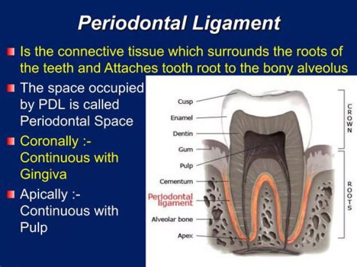
Like all soft fibrous connective tissues, the periodontal ligament consists of a fibrous stroma in a gel of ground substance containing cells, blood vessels and nerves. The fibrous stroma consists primarily of collagen (with very small amounts of oxytalan) and the cells are mainly fibroblasts (Fig. 1).
Do all teeth have periodontal ligaments?
The periodontal ligament is only found between the tooth root and adjacent bone and does not support the outer gum tissues. The complex nature of the PDL tissue allows the tooth to properly function during chewing and to withstand the pressure from grinding or clenching.
What does a periodontal ligament do?
Periodontal Ligament Stem Cells (PDLSCs) The Periodontal Ligament (PDL) can be defined as a fibrous joint that anchors the root of the tooth to the alveolar bone socket. Made of fibrous connective tissue, it holds the tooth in sprung suspension enabling the tooth to take on the mechanical pressure during function.
Where is the periodontal ligament found?
What causes widening of the periodontal ligament?
PDL widening occurs in trauma from occlusion, but in association with angular bone defects and mobility of teeth. However, in scleroderma, involved teeth are often not mobile and their gingival attachments are usually intact.
Why is the periodontal ligament important?
By all means, a primary purpose of the periodontal ligament was to provide the network of connective tissue fibers that connect the boney socket to the cementum on the root surface. It also acts as a cushion, a shock absorber of sorts to protect the tooth and the jaw bone from the trauma of chewing.
What is the periodontal ligament capable of?
The periodontal ligament holds the teeth in sprung suspension, with the result that each tooth is capable of small movements in its alveolar bone socket. Blood vessels and nerves are also found at the junction between the dental root and alveolar bone.
What are the radiographic signs and symptoms of periodontal disease?
7 Moderate Marginal Periodontitis Generalized form demonstrates horizontal bone loss Localized defects include vertical bone loss and loss of buccal and lingual cortices Loss of buccal or lingual cortex is difficult to view radiographically. It may be seen as decreased density over the root surface
How does the periodontal ligament ( PDL ) affect a tooth?
The ligament can enlarge and allow the tooth to become loose. Once the excessive forces on the tooth are reduced, the PDL will heal, and tooth mobility will decrease.
Where is the center of rotation of the periodontal ligament?
The periodontal ligament allows movement around a center of rotation. The center of rotation is midroot. Therefore, the greatest movement will be at the apex and alveolar crest. As bone is lost, the center of rotation moves toward the apex </li></ul>
How is bone loss camouflaged by periodontal disease?
Any minor bone loss on the buccal aspect of the tooth may be overlapped by the intact lingual bone, thus bone loss on one aspect of tooth is camouflaged by bone on the opposite side. Hence, the early signs of periodontitis such as deepening of periodontal pocket or recession are best visualized clinically.
